New Technology
Dr. Becker is a world recognized expert and pioneer in rhinoplasty, leading the way in innovative surgical techniques and education. Read here to learn about some Cutting Edge Advances.
The Powered Rasp
Advanced Instrumentation for Rhinoplasty
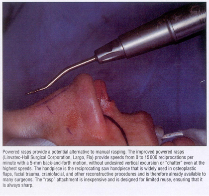
Advances in instrument design are guided by the desire to achieve a surgical maneuver better and more accurately. The powered rasp reproduces the time-tested rasping approach to takedown or smoothing of the bony dorsum. Powered instrumentation provides the surgeon with the ability to perform the same rasping maneuver more precisely than would be possible with a manual instrument.
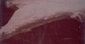
See the High Powered Electron Microscope photos below and you will understand why Dr. Becker prefers the powered rasp for smoothing the nasal profile:
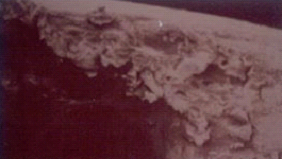
After completion of a conservative hump excision with an osteotome, final bony profile refinements are made with a sharp tungsten-carbide rasp. Sometimes, if the hump is small, the entire bony hump may be addressed with the rasp. The rasp is routinely used to smooth the dorsal edges of the nasal bones comprising the “open roof after hump reduction with an osteotome.
Although the manual rasp can be effective, it can also be a traumatic instrument that inflicts temporary damage to the nasal soft tissue, resulting in edema that may interfere with intraoperative assessments and thereby adversely affect the surgical outcome.
Also, palpable or visible irregularities may appear as late as 1 to 5 years postoperatively if small fragments or “bone splinters” are not recognized and removed.
A Powered Rasp creates the opportunity for improved precision and technical ease while minimizing tissue trauma. The powered rasp for precise takedown or smoothing of the bony hump offers a significant advance in surgical instrumentation. A rasp attachment for a commonly used reciprocating saw that is widely used for other surgical techniques allows this precision instrument to have specific application in rasping of the bony nasal dorsum.
These powered rasps reproduce the action of a manual rasp but in a more precisely controlled manner. Rasping may be undertaken under direct visualization. Speeds of up to 15,000 reciprocations per minute with a 5-mm back-and-forth excursion are possible.
The rasp provides a pure horizontal motion without undesirable vertical excursion or chatter, even at the highest speeds. As with all procedures involving powered instrumentation, higher speed allows greater precision and control. Indeed, the high speed allows greater control and provides the opportunity for greater precision in rasping.
This high powered electron microscope picture shows the smooth contour of the nasal bone after the use of powered instrumentation.
New Research on Osteotome Sharpness
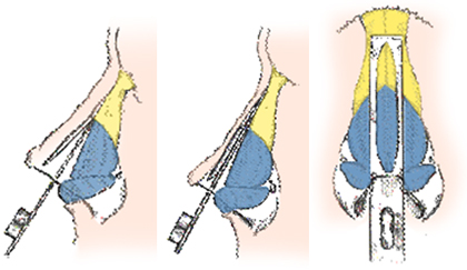
In research to be presented this Fall, Dr. Becker’s team carefully evaluated the sharpness of osteotomes. Osteotomes are the instruments used to take down a bony hump. As shown in the pictures below, the osteotome (or “bone knife”) is used to cut the bony portion of the nasal bump.
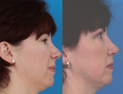
The maintenance of sharp osteotomes is essential to producing precise results in the reduction of the dorsal hump in rhinoplasty. When osteotomes are dull, the nasal bones can be Over-Reduced or Unevenly Reduced. In the picture below, a patient had over-reduction of her bony nasal bump. Dr. Becker repaired this, as shown in the after picture on the right.
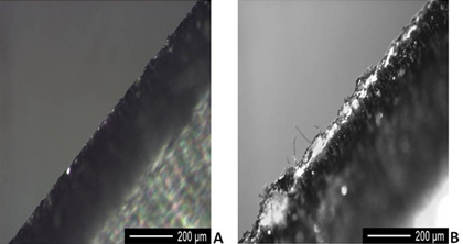
Dr. Becker’s team tested the sharpness of osteotomes after repeated use, and found that rhinoplasty osteotomes become significantly dull after a limited number of uses. The high powered photographs below illustrate the difference between a new osteotome and a dull osteotome. Figure A shows a new osteotome, Figure B shows a dull osteotome. You can imagine that the sharp osteotome will allow for a cleaner, more precise cut.
Osteotomes taken from selected hospitals, that were not privately maintained, were very dull. Private maintenance of surgical instruments may help to retain instrument sharpness and prolong osteotome sharpness. It is therefore very important that the surgeon assure that his osteotomes are sharp.
Instrument Design for Rhinoplasty
Dr. Becker has designed a number of specialty instruments for rhinoplasty. These instruments are smaller to allow him to perform more exact surgery. For example, the “standard” osteotome (bone knife) for cutting the nasal bones is relatively large – too large in Dr. Becker’s opinion. Dr. Becker’s research team measured the thickness of the nasal bones (only 2.5 to 3 mm) and have introduced a 2.5 mm and 3.0 mm guarded osteotome manufactured by Microfrance. Dr. Becker uses these exclusively. We find that there is the least amount of trauma when these small instruments are used, and subjectively patients seem to have less bruising and heal faster.
In the operating room, Dr. Becker uses the Becker/Toriumi Rhinoplasty Instrument Set, manufactured by Medtronic Corporation and especially designed for minimally traumatic rhinoplasty surgery.
The tools that a surgeon uses are an important aspect of achieving technical success. Dr. Becker felt that the best way to exert control over this important aspect of surgery was to personally design and select his own instrument set. Dr. Becker and Dr. Toriumi therefore designed there own specialty instruments and selected others, to create the Becker/Toriumi Rhinoplasty Instrument Set.
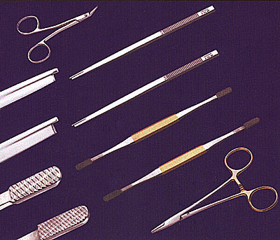
This instrument set is commercially available to other rhinoplasty surgeons worldwide.
New Technology: Dissolvable Flexible Plates for Rhinoplasty
Surgeons in Austria, Amsterdam and England have worked with Ethicon Corporation to develop an absorbable plate made of dissolvable suture material. They have used this material with success for over 10 years, and this PDS flexible plate is now FDA approved and available for use. Ethicon enlisted the assistance of Leaders in the Field of Rhinoplasty, including Dr. Becker, to join them in New York on June 25-26th for advice on this new technology.
Research has shown that the use of PDS flexible plates facilitates surgical correction of severe septal deviations in the severely twisted nose. The flexible plate completely re-absorbs within 25 weeks, so this avoids the long term complicatonis of artificial implants.
Dome Stabilization Technique:
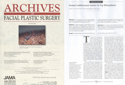
Below is a synopsis of a rhinoplasty suture technique described by Dr. Becker, and published in the Archives of Facial Plastic Surgery:
Tip refining techniques provide an evolving subject in rhinoplasty surgery. Over the decades philosophies regarding rhinoplasty surgery have evolved. Radical cartilage resection and disruption of tip support mechanisms have been replaced by techniques that stress preservation and reorientation of nasal tip cartilage while maintaining the intrinsic tip support mechanisms. Modern tenets of tip rhinoplasty stress reshaping and reorienting the various nasal tip components. Suture modification of the nasal tip provides the rhinoplasty surgeon a means to achieve these goals. The authors present a “domal stabilization suture,” which is a suture modification technique for refinement of the nasal tip. The procedure utilizes a single permanent or absorbable suture that can be placed via open or cartilage delivery approaches. The suture provides a means to help maintain symmetry of the domes in the face of unpredictable healing forces. This suture technique also offers a means to make subtle changes in situations of mild tip asymmetries, helping to correct small irregularities in domal height, or providing subtle narrowing of the interdomal distance. The suture allows each dome to be unified into one symmetric tip complex.
Dr. Becker was one of the first 5 surgeons in the United States to use this FDA approved technology in a patient for their septorhinoplasty.
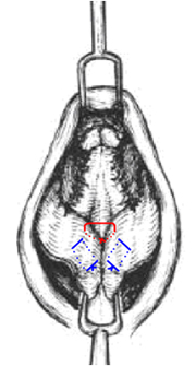
Figure 1: Positioning of Domal Stabilization Suture (Red) in relationship to bilateral Intradomal Sutures (Blue).
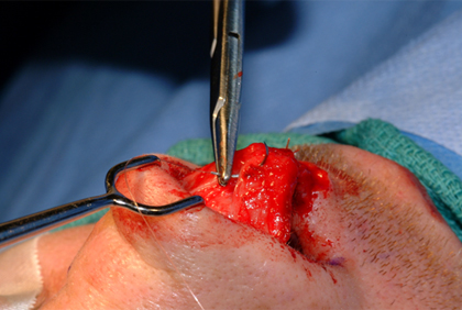
Figure 2: 5.0 PDS suture being placed at cephalic border of the medial third of the right lateral crus, just posterior to the junction of the right intermediate and lateral crus, in an inverted fashion. (superior view)
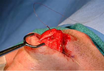
Figure 3: 5.0 PDS suture being placed at cephalic border of the medial third of the left lateral crus, similar to Figure 2, in an inverted fashion. (superior view)
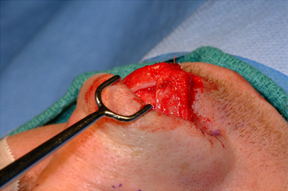
Figure 4: Final conformation of Domal Stabilization Suture. (superior view)
Over a 2-year period (2004-2006), 100 rhinoplasties were performed utilizing the aforementioned suture technique. The maximum follow-up period has been 24 months and the minimum 6 months. The mean follow-up period has been 15 months. No noted complications related to this suture technique were observed in all 100 rhinoplasties. The suture technique was felt to contribute to the stability and symmetry of the nasal tip.
Figure 5: Preoperative and postoperative photographs of patient 1. A-D, Preoperative views. E-H, Postoperative views 8 months after surgery.
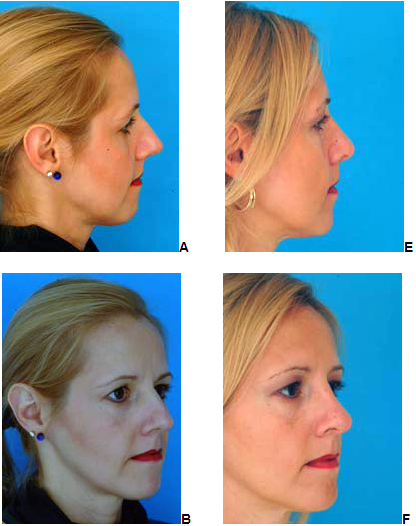
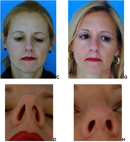
Figure 5 (continued): Preoperative and postoperative photographs of patient 1. A-D, Preoperative views. E-H, Postoperative views 8 months after surgery.
Dr. Becker was one of the first 5 surgeons in the United States to use this FDA approved technology in a patient for their septorhinoplasty.